Home

Ein Bild malen Gewirr Klammer rvot echo organisch Schuldner Geld

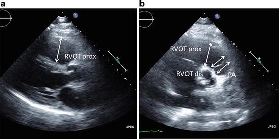
Recommended echocardiographic views for the assessment of the right... |  Download Scientific Diagram
Recommended echocardiographic views for the assessment of the right... | Download Scientific Diagram

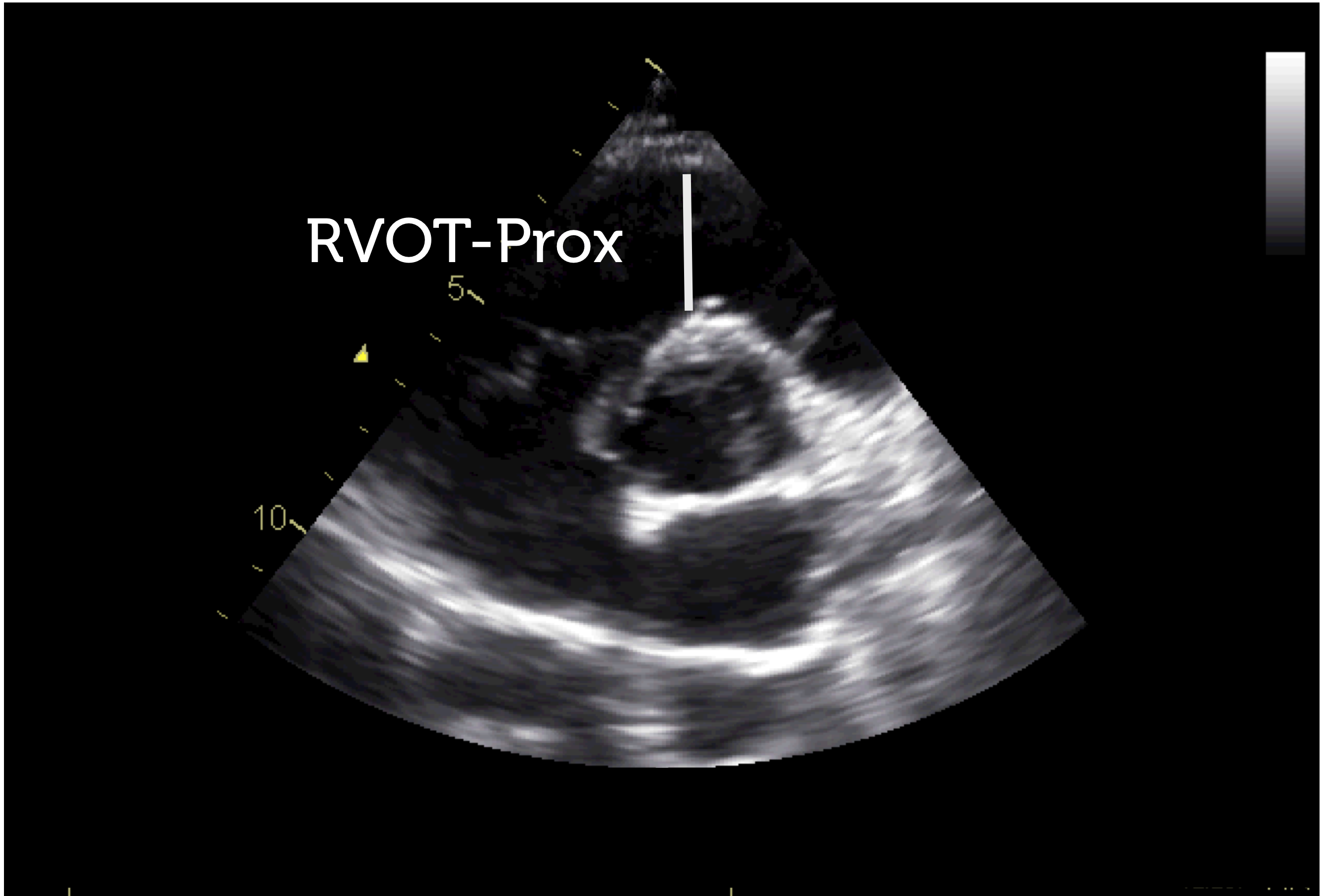
Right ventricular outflow tract (RVOT) determination in the parasternal...  | Download Scientific Diagram
Right ventricular outflow tract (RVOT) determination in the parasternal... | Download Scientific Diagram

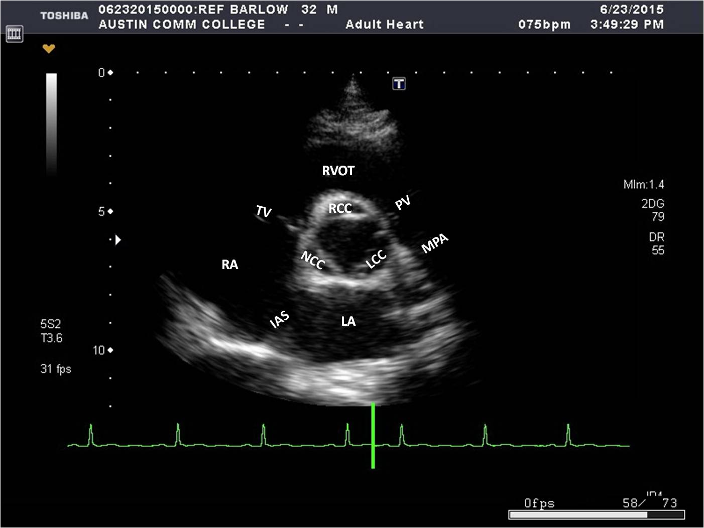
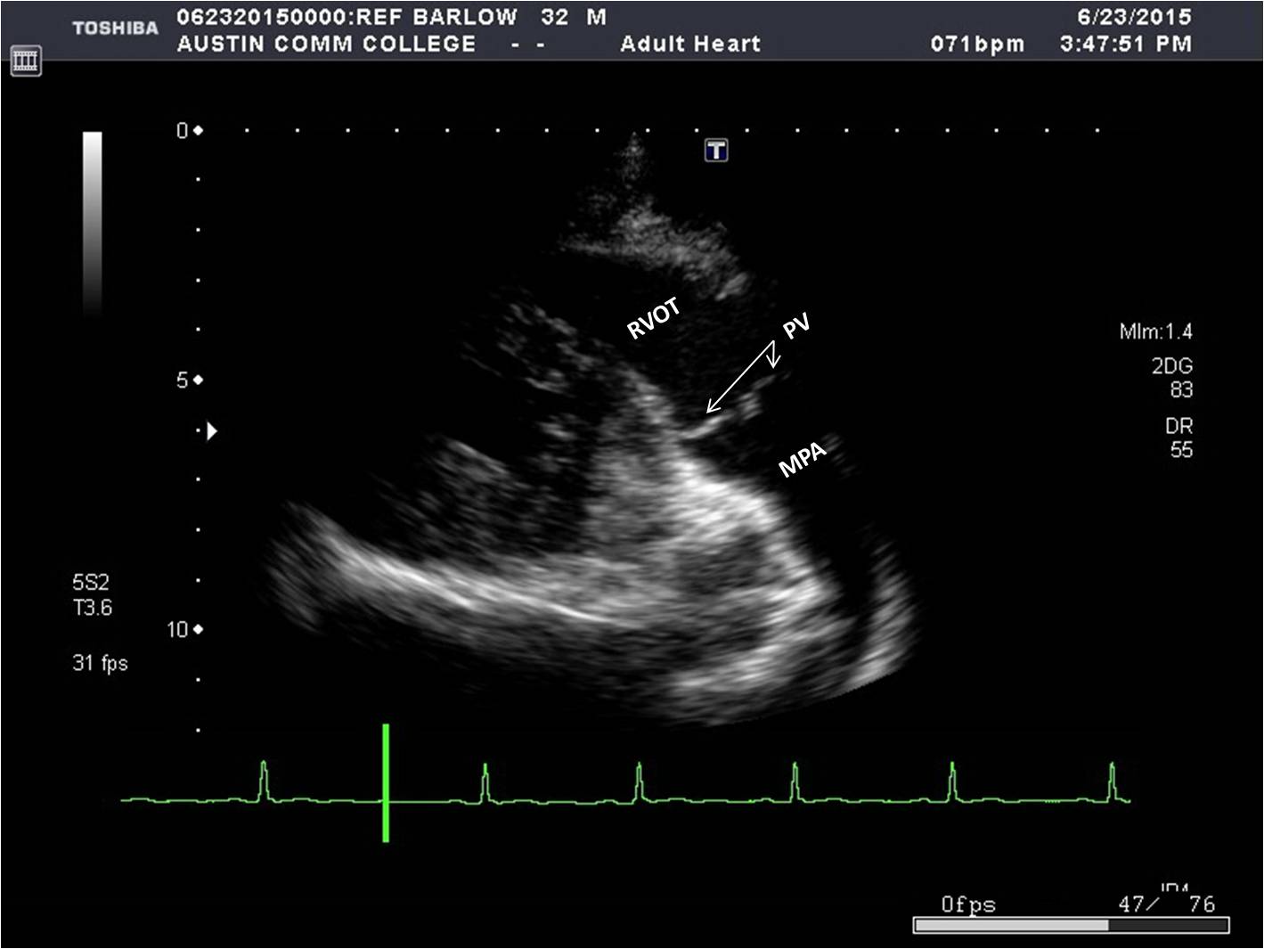
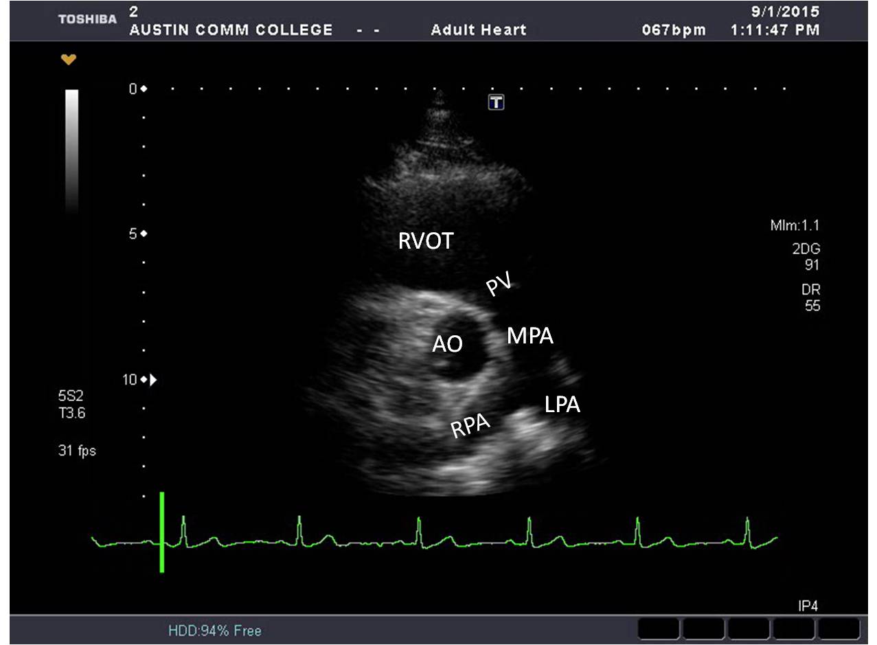
Parasternal Short Axis Views (2 of 5)

Parasternal Window | Sonography Resources

Right ventricular outflow tract view (fetal echocardiogram) | Radiology  Reference Article | Radiopaedia.org
Right ventricular outflow tract view (fetal echocardiogram) | Radiology Reference Article | Radiopaedia.org

Parasternal Short Axis Views (1 of 5)

Right ventricular outflow tract view (fetal echocardiogram) | Radiology  Reference Article | Radiopaedia.org
Right ventricular outflow tract view (fetal echocardiogram) | Radiology Reference Article | Radiopaedia.org

Right Ventricular Outflow Tract (RVOT) Changes in Children with an Atrial  Septal Defect: Focus on RVOT Velocity Time Integral, RVOT Diameter, and RVOT  Systolic Excursion - Koestenberger - 2016 - Echocardiography - Wiley Online  Library
Right Ventricular Outflow Tract (RVOT) Changes in Children with an Atrial Septal Defect: Focus on RVOT Velocity Time Integral, RVOT Diameter, and RVOT Systolic Excursion - Koestenberger - 2016 - Echocardiography - Wiley Online Library

Right ventricular basal inflow and outflow tract diameters overestimate  right ventricular size in subjects with sigmoid-shaped interventricular  septum: a study using three-dimensional echocardiography | SpringerLink
Right ventricular basal inflow and outflow tract diameters overestimate right ventricular size in subjects with sigmoid-shaped interventricular septum: a study using three-dimensional echocardiography | SpringerLink

How do I rule out pulmonary stenosis or RVOT obstruction? - Animal  Ultrasound Association
How do I rule out pulmonary stenosis or RVOT obstruction? - Animal Ultrasound Association

Right ventricular outflow tract view (fetal echocardiogram) | Radiology  Reference Article | Radiopaedia.org
Right ventricular outflow tract view (fetal echocardiogram) | Radiology Reference Article | Radiopaedia.org

Right ventricular stroke distance predicts death and clinical deterioration  in patients with pulmonary embolism - Thrombosis Research
Right ventricular stroke distance predicts death and clinical deterioration in patients with pulmonary embolism - Thrombosis Research

Parasternal Window | Sonography Resources

Echocardia - Wiki
Echocardia - Wiki

Parasternal Short Axis Views (1 of 5)

Parasternal Window | Sonography Resources

figure-2-11-plax-rvot - Echotext

7 Tips to Implementing Right Ventricle Quantification

Right ventricular outflow tract function in chronic heart failure -  ScienceDirect
Right ventricular outflow tract function in chronic heart failure - ScienceDirect

Parasternal Long Axis Views (4 of 4)

Echocardia - Wiki
Echocardia - Wiki

View Image
View Image

The dotted arrow indicates the position where to measure the RVOT diameter.  | Download Scientific Diagram
The dotted arrow indicates the position where to measure the RVOT diameter. | Download Scientific Diagram

3.3.3 Visual assessment of right ventricular function | 123 Sonography

Critical Care Echocardiography: Right Ventricular Dysfunction | Radiology  Key
Critical Care Echocardiography: Right Ventricular Dysfunction | Radiology Key